Ouch, they hurt!! Kidney stone is no fun.
BY Dr. Sudhir Thaduri Published on August 18, 2021
Kidney stones have a reputation for being very painful such that the severity of pain is often compared to labour pain in women. They are pretty dramatic and can take someone by surprise. Kidney stones are a fairly common occurrence among seemingly healthy individuals. Most people would know of someone in their friends or family visiting Emergency for kidney stone pain. For many, it may be a one-time event with a happy ending after the stone gets passed, while for others, it may need a surgical procedure to extract the stone or keep forming stones recurrently.
Types of Stones:
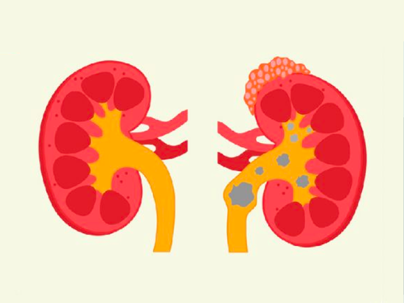
Most stones (nearly 2/3rds) are chemically composed of Calcium oxalate or Calcium phosphate, while the less common ones include uric acid, struvite, or cysteine stones. Stones are believed to result from an excessive concentration of stone-forming material in the urine due to dehydration or innate abnormality of the kidney in regulating factors that would normally avoid precipitation of stone material.
Symptoms:
Up to a 3rd of stones may not cause any symptoms and are detected incidentally during abdominal imaging done for other purposes. Of these, about a 3rd, in turn, end up having symptoms sooner or later. Pain is the most common symptom that results from the movement of stone starting from the kidney enroute to the urinary bladder with a corresponding location that changes with the descent of the stone, often coinciding with painful paroxysms as the ureteric contractions try to expel the stone. If the stone is small (<5mm) and not impacted, the body may successfully expel the stone in urine with relief of pain. Alternatively, it may get impacted and result in obstruction to urine resulting in backpressure on the kidney with impairment of function by damming up the urine flow. Patients may be appalled, have vomiting, or have radiation of pain to the groin area. Rarely if the obstructed urinary tract gets infected, it could result in fever, chills, and sick patients. Blood in the urine is a common accompanying symptom that could be visible to the eye or microscopic.Individuals who have a tendency to form stones recurrently or form stones bilaterally could endure obstruction to one or both sides, especially in stones formed of Struvite material that causes progressive loss of kidney function on one or both sides resulting in the need for dialysis or transplantation. Fortunately, kidney stones are very rare as a cause of permanent kidney damage, and less than 5% of individuals with kidney stones end up in End-stage kidney disease.
Diagnostic Evaluation: When suspected of kidney stone, initial testing includes a Non-contrast CT scan of the abdomen, which is the best diagnosis modality. An ultrasound of kidneys could be performed for pregnant women or if CT is not available. Intravenous pyelography and MRI are alternate modalities of imaging that can be used if CT or ultrasound is not available. CT is very sensitive in picking up stones 2mm or greater and delineates any urinary tract obstruction. CT can also indicate the type of stone based on its density and identify specific patterns in rare genetic conditions of recurrent stone formation like Medullary Sponge Kidney Disease.
Other conditions that may mimic kidney stone pain and need to be excluded on evaluation for acute pain may arise from other structures in the abdominal cavity: Rupture/torsion of ovarian cyst, ectopic pregnancy, dysmenorrhea, appendicitis, diverticulitis, cholecystitis, mesenteric ischemia, pyelonephritis, or rarely from shingles of abdominal skin.
Management: Most patients can be treated as outpatients with pain control and hydration, anticipating that the stone would pass on its own. However, large stones that get obstructed or severe symptoms that preclude oral medicines or complications like infection may need hospital admission and urgent referral to a Urologist.
Stone passage: Depends on size and location. Most stones <5mm pass by themselves, and Stones>10mm usually need surgical removal. As the stone gets bigger, there is an increased need for urologic intervention. Most stones tend to pass within 4-6 weeks.
Medical expulsive therapy: Medications (Alpha-blockers and calcium channel blockers) that relax the ureter and facilitate stones can help the passage of stones between 5-10mm within a few weeks. If stones are not passed within 3-4 weeks, a repeat CT scan or X-rays can be obtained, and patients are usually referred at that point to a Urologist for intervention. Other reasons to seek Urologist consultation would be for large stones, bilateral stones obstructing the outflow of urine, or unrelenting symptoms. Urologists have various options depending on location/proximity of stones, including laser lithotripsy, percutaneous stone removal, or may rarely need laparoscopic stone removal. In rare situations with severe infection and obstruction of urine flow, tubes can be placed directly from the flank skin into the collecting system of the kidneys to drain and relieve pressure in an obstructed system (Percutaneous nephrostomy).
Prevention:
Most people who get a kidney stone may never get another one, but quite a few of them continue to form recurrent stones and need evaluation with a 24hr urine testing to look at specific abnormalities in the urinary solute, which lead to precipitation and stone formation. An underlying cause of stone can be found in most instances by urine analysis and by stone analysis if the stone is collected using a strainer by the patient or removed by surgical procedure. Specific medicines may be given to target urinary abnormalities depending on the type of stones. In addition to specific therapy, general preventive measures applicable to any form of stone formers include optimal hydration (up to 2 litters of urine per day), or patients could watch their urine stay clear, which is a surrogate for adequate urinary dilution to prevent precipitation of stone material in the urinary tract. General advice to reduce/moderate intake of dietary sodium, sugars, and animal protein help ameliorate all types of stones by reducing the formation of the nidus, which serves as the seed for stone growth. Increasing dietary potassium through fruits and vegetables reduces the common type of stones formed from calcium oxalate. All stone formers tend to benefit from weight loss. Specific dietary restrictions may be needed based on stone constituents like oxalate, uric acid, cysteine to help prevent their increased concentration in the urinary tract leading to stone formation. Some stones that can assume huge size like the “struvite” or “stag horn” may need antibiotic therapy to sterilize ammonia generating bacteria that can form stones rapidly.
To conclude, kidney stones are a common cause of acute abdominal pain which are quite dramatic in presentation and can mimic other serious conditions that need to be differentiated from it. Understanding that stones can be prevented to a large extent by optimal hydration and paying heed to a diet low in sodium, animal protein, and sugars can go a long way in avoiding this painful condition.

MBBS, MD, DNB, Assistant Professor, Division of Nephrology, Transplant, University of Birmingham Alabama.

In the age of digitalization, online consultation with a doctor has become an increasingly popular option for people seeking medical advice. But how does online consultation compare to traditional in-person doctor's visits?

Teleconsultation is a viable option for people who need mental health care but are unable to access it in person. In this blog post, we will explore the benefits of teleconsultation for mental health care and how it can help people manage their mental health.

In this blog post, we will discuss the features and benefits of various teleconsultation apps and provide recommendations for choosing the right one for your needs.